
Gastroshiza is a serious yet treatable congenital anomaly that affects the abdominal wall of a developing fetus. It is typically identified before birth through prenatal imaging and occurs when the baby’s intestines protrude outside of the body through an opening near the belly button, without a protective sac. This condition can seem alarming to expecting parents, but with prompt medical intervention, including surgical repair shortly after birth, the outcomes for infants with Gastroshiza have significantly improved over the years.
In this article, we will explore the comprehensive details about Gastroshiza including its definition, causes, signs and symptoms, diagnostic methods, treatment options, post-surgery care, long-term outlook, and more. This guide aims to provide a thorough understanding of gastroschisis in clear, non-technical terms suitable for both parents and healthcare professionals seeking deeper insights.
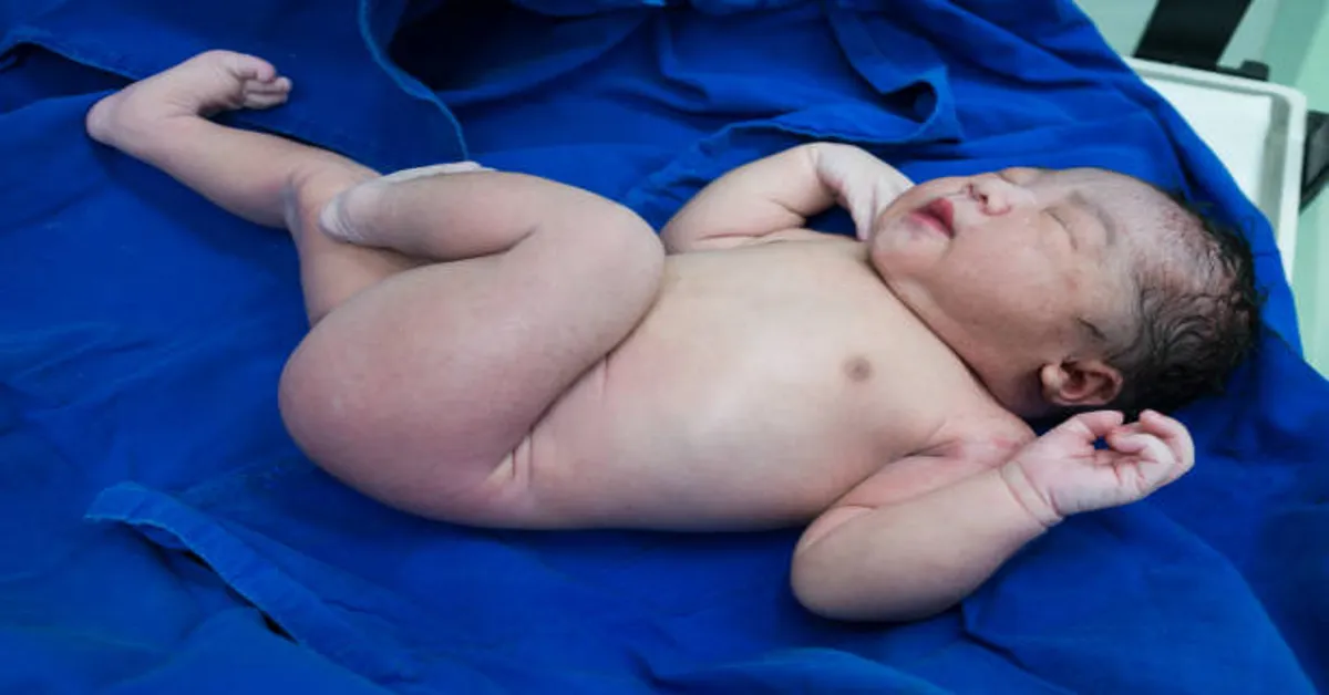
What Is Gastroshiza?
Gastroshizais a congenital (present at birth) defect of the abdominal wall. It typically presents as an opening located to the right of the umbilicus (belly button), through which the baby’s intestines and occasionally other organs such as the stomach or liver protrude outside the body. Unlike another similar condition called omphalocele, in Gastroshiza, the herniated organs are not enclosed in a membrane or sac. This exposes them directly to amniotic fluid during pregnancy, which can cause inflammation and thickening of the intestines.
It is a structural defect and not related to genetic syndromes in most cases. The condition is usually isolated, meaning the baby does not have other major congenital abnormalities, although complications can arise if not managed properly.
Causes and Risk Factors
The exact cause of Gastroshizaremains unknown. However, medical researchers believe it stems from a vascular disruption during fetal development, particularly involving the right omphalomesenteric artery. This disruption leads to impaired development of the abdominal wall and allows intestines to protrude through the defect.
Although there is no definitive genetic or inherited cause, certain risk factors have been identified that may increase the likelihood of Gastroshiza:
Maternal Age
Young maternal age, especially teenagers and women under 20, is strongly associated with a higher risk of having a baby with Gastroshiza. The exact reason for this association is not clearly understood, but it is one of the most consistent epidemiological findings.
Smoking and Substance Use
Maternal smoking, use of illicit drugs, or alcohol during pregnancy may contribute to fetal vascular disruptions or developmental anomalies, including Gastroshiza.
Environmental Exposures
Exposure to certain agricultural chemicals, such as herbicides or pesticides, has been proposed as a potential contributing factor in some studies.
Nutritional Deficiencies
Inadequate intake of folic acid or poor overall maternal nutrition may affect fetal development and increase the risk of Gastroshiza.
Infections and Inflammatory Factors
Some infections or inflammatory processes during early pregnancy may interfere with normal development of the abdominal wall, though this is less commonly implicated.
Signs and Symptoms
Because Gastroshiza is a physical defect visible at or before birth, the condition is usually detected during routine prenatal care. However, the signs and symptoms can be divided into prenatal and postnatal presentations.
Prenatal Signs (During Pregnancy)
- Ultrasound Findings: The most common sign is visualization of free-floating bowel loops outside the fetal abdomen during routine ultrasound, typically around 18 to 20 weeks gestation.
- Abnormal Maternal Serum AFP: Alpha-fetoprotein (AFP) levels are often elevated in mothers carrying fetuses with abdominal wall defects, prompting further investigation.
Postnatal Symptoms (After Birth)
- Visible Intestinal Protrusion: At birth, the intestines are visible outside the body, typically without a covering sac, and are often red and swollen due to exposure to amniotic fluid.
- Feeding Intolerance: The infant may not tolerate oral feeding until bowel function returns post-surgery.
- Intestinal Dysfunction: Bowel movements may be delayed, and the baby may show signs of poor digestion initially.
Diagnosis
Prenatal Diagnosis
Early detection is crucial for planning delivery and postnatal care. Diagnosis typically occurs during the second trimester through:
- Ultrasound Examination: The gold standard in detecting gastroschisis, showing free-floating intestines outside the fetal abdomen.
- Maternal Blood Tests: High levels of alpha-fetoprotein (AFP) can indicate an abdominal wall defect but are not specific to gastroschisis.
Postnatal Diagnosis
At birth, visual confirmation is straightforward. A complete physical examination and imaging tests such as X-rays or abdominal ultrasound may be used to assess the extent of intestinal involvement and rule out other anomalies.
Treatment and Surgical Intervention
The cornerstone of gastroschisis treatment is surgical repair, typically carried out shortly after birth. The primary goals of treatment are to:
- Protect the exposed intestines
- Return the organs to the abdominal cavity
- Close the abdominal wall defect
Immediate Postnatal Care
Once the baby is born, immediate steps are taken to reduce the risk of infection, fluid loss, and further injury to the exposed organs:
- The infant is placed in a sterile, warm environment.
- The exposed bowel is covered with sterile material or a plastic silo bag.
- IV fluids and antibiotics are started to prevent dehydration and sepsis.
- A nasogastric tube is inserted to decompress the stomach.
Surgical Repair Approaches
Primary Repair
In cases where the bowel is healthy and the abdominal cavity can accommodate the intestines, the surgeon performs a single surgery to place the organs back inside and close the defect.
Staged Repair (Silo Method)
If the intestine is too swollen or the abdominal cavity is too small, a silo (a sterile plastic pouch) is used to gradually push the intestines back inside over several days. Once the organs are returned, the abdominal wall is surgically closed.
Postoperative Care and Recovery
After surgical repair, infants require monitoring in a neonatal intensive care unit (NICU). Postoperative care includes:
- Parenteral Nutrition (IV Feeding): As the bowel needs time to recover, infants may receive nutrition through an IV for several days to weeks.
- Gradual Introduction of Oral Feeds: As bowel function returns, feeding is slowly introduced through a feeding tube or bottle.
- Pain Management: Pain control is managed with appropriate medications.
- Monitoring for Complications: These may include infection, bowel obstruction, or poor wound healing.
Full recovery can take several weeks depending on the severity of the defect, the condition of the intestines, and the presence of any complications.
Long-Term Outlook and Complications
The long-term prognosis for babies with gastroschisis has improved significantly due to advances in neonatal care and surgical techniques. Most babies recover fully and lead healthy lives. However, some may face challenges depending on the severity of the condition and surgical outcomes.
Possible Complications
- Short Bowel Syndrome: If large portions of the intestine are damaged or removed, the baby may have difficulty absorbing nutrients.
- Intestinal Adhesions: Scar tissue may form, causing future bowel obstruction or digestive issues.
- Feeding Difficulties: Some babies may experience delayed feeding or food intolerance.
- Growth Delay: Infants may require nutritional support to ensure normal growth and development.
Follow-Up Care
Babies with gastroschisis typically require ongoing pediatric care to monitor:
- Nutritional status and weight gain
- Gastrointestinal function
- Developmental milestones
- Potential surgical complications
Prevention and Prenatal Management
Because the exact cause of gastroschisis is unknown, there are no guaranteed ways to prevent it. However, certain preventive steps and prenatal strategies may reduce the risk or improve outcomes:
- Early Prenatal Care: Regular check-ups help monitor fetal development and detect anomalies early.
- Healthy Lifestyle Choices: Avoiding smoking, drugs, and alcohol during pregnancy reduces the risk of many congenital conditions.
- Nutritional Supplements: Folic acid and prenatal vitamins may support fetal health.
- Planned Delivery: In high-risk pregnancies, a planned delivery at a tertiary care center ensures immediate access to neonatal surgery and intensive care.
Emotional Support for Families
A diagnosis of gastroschisis can be emotionally overwhelming for parents. It is essential that families receive compassionate support, accurate information, and access to counseling when needed. Support groups, mental health professionals, and genetic counseling can all be valuable components of family care during and after pregnancy.
Conclusion
Gastroschisis is a serious but manageable congenital condition involving a defect in the abdominal wall through which the intestines protrude. Early detection through prenatal screening and coordinated care between obstetricians, neonatologists, and pediatric surgeons can lead to excellent outcomes. With timely surgical intervention, careful postoperative care, and continued monitoring, most infants born with gastroschisis go on to thrive. While the initial diagnosis may be daunting, modern medical advances and support systems offer hope and healing for affected families.
ALSO READ: Blazertje: The Essential Guide to Modern Blazer Fashion
Frequently Asked Questions (FAQs)
1. Is gastroschisis a genetic disorder?
No, gastroschisis is not typically considered a genetic or inherited condition. It usually occurs sporadically and is not associated with chromosomal abnormalities in most cases.
2. How early can gastroschisis be detected during pregnancy?
Gastroschisis can usually be detected during a routine second-trimester ultrasound, often between 18 to 20 weeks of gestation.
3. Can a baby with gastroschisis be delivered vaginally?
Yes, many babies with gastroschisis can be delivered vaginally unless there are other complications that require a cesarean section. The delivery plan is made in consultation with the obstetrics and neonatal teams.
4. Will my baby need multiple surgeries for gastroschisis?
Not all babies need multiple surgeries. If a primary repair is possible, only one surgery may be needed. In more complex cases, staged surgeries may be required.
5. What is the long-term outlook for children born with gastroschisis?
Most children recover well after surgical repair and grow up healthy, although some may have ongoing digestive or nutritional needs that require monitoring and support.